We follow the guidelines set by DFMS and ISUOG!
What are 2D and 3D/4D scans?
A 2D scan shows two-dimensional images of the fetus. 2D scans are very suitable for fetal diagnostics because you look ‘into’ the body. We start all scans with a 2D scan. In a 3D scan, you can see three-dimensional images of the fetus’s own unique features. 4D scans mean watching 3D images in motion, like a movie. For example, you can see your baby yawn, smile, open it’s eyes or suck on a thumb. You can do 3D/4D throughout your pregnancy. However, the fetus gets some subcutaneous fat in the 2nd trimester – and with this its personal features. After week 32 the amniotic fluid is displaced due to the size of the fetus and the image quality deteriorates. Therefore the best time for 3D/4D scans is between weeks 26 and 32 (for twins between weeks 23-28). This is the time where the amount of amniotic fluid tops and the head has still not set in your pelvis. If you only want to be scanned to see how your baby looks, we recommend that you book an appointment during this period.
What is ultrasound?
Ultrasound is high frequency sound waves that can not be captured by the human ear. During ultrasound examination a transducer is put against your belly. The sound waves are now transmitted from the transducer to the fetus, causing an echo to be thrown back and captured, again by the transducer. The echo translates into detailed images of the fetus and uterus, which can be seen and analyzed on a screen. In a doppler study, we can see the blood flow in the fetus and umbilical cord and we can measure the resistance in the vessels. It is considered harmless for a pregnant woman and the fetus to conduct ultrasound examinations. Our attitude, however, is that you should be trained and experienced in fetal diagnostics and at Spire – of course – we all are!

Limitations to ultrasound
There may be cases where the fetus is positioned in a way so that the visibility is limited. In these cases we may nudge your baby to move by giving your belly a small push. You may also be asked to stand up or move a little to get the fetus to change position. The fetus is safely surrounded by tissue and amniotic fluid and will not be harmed by this procedure.
It is very important for the image quality that there is an amount of fluid in front of the part of the fetus that we want to investigate. If the amniotic fluid has decreased or if the fetus is right against the uterine wall or otherwise ‘hides’, the visibility may be impaired. The same applies if there are fibromas in the uterine wall or if the mother has a BMI higher than 25 but in most cases the scan turns out fine anyway!
When you study a fetus with ultrasound you get lots of valuable information about growth, health and possibly anomalities. Ultrasound, however, is never a guarantee that the fetus is a hundred percent healthy.

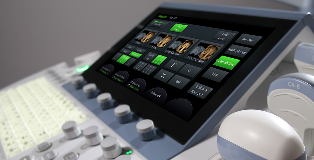
At Spire you will meet a professional team of sonographers. We do all types of pregnancy scans including 3D and 4D. We have the latest equipment ensuring great looking images with all scans.
We are registered with the Danish Patient Safety Authority.
Follow us

